
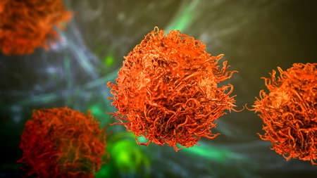
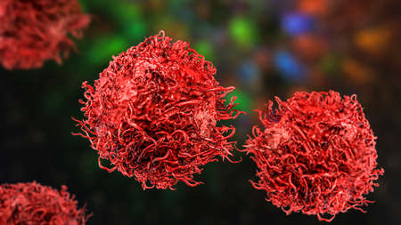
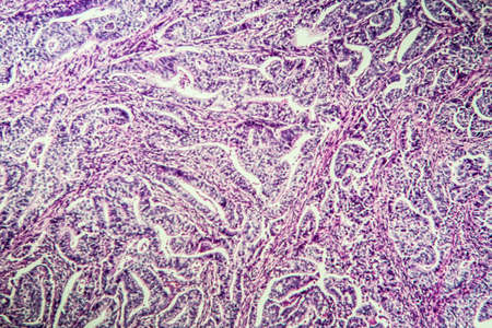
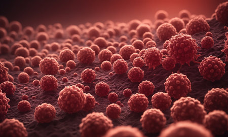
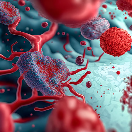
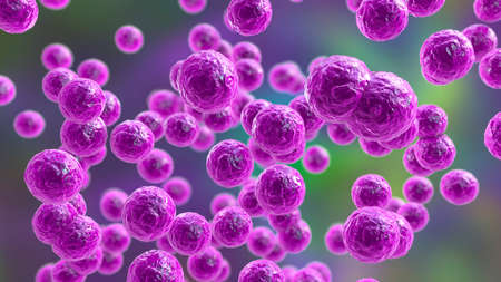
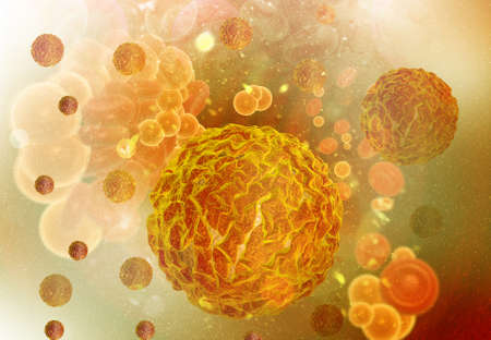
Stomach cancer cells, 3D illustration showing morphology of cancerous cells
Коллекция по умолчанию
Коллекция по умолчанию
Создать новую
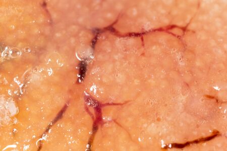
fish caviar as a background. macro
Коллекция по умолчанию
Коллекция по умолчанию
Создать новую
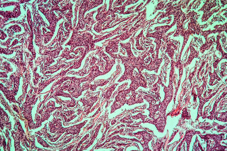
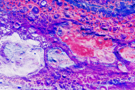
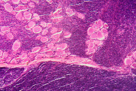
Breast cancer of the woman diseased tissue 100x
Коллекция по умолчанию
Коллекция по умолчанию
Создать новую
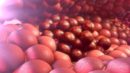
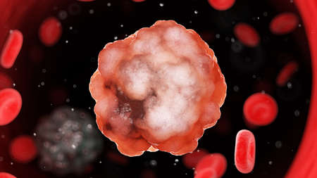
Cancer cells
Коллекция по умолчанию
Коллекция по умолчанию
Создать новую
Stomach cancer cells, 3D illustration showing morphology of cancerous cells
Коллекция по умолчанию
Коллекция по умолчанию
Создать новую
3d rendered illustration of a cancer cell
Коллекция по умолчанию
Коллекция по умолчанию
Создать новую
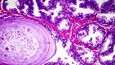
Histopathology of prostate gland hyperplasia, light micrograph, photo under microscope
Коллекция по умолчанию
Коллекция по умолчанию
Создать новую
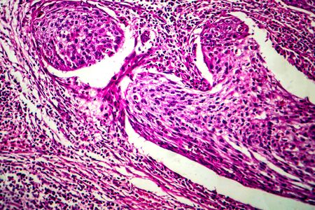
Squamous cell carcinoma of the uterus, light micrograph, photo under microscope
Коллекция по умолчанию
Коллекция по умолчанию
Создать новую

Cancer cell, malignant tumor cell
Коллекция по умолчанию
Коллекция по умолчанию
Создать новую
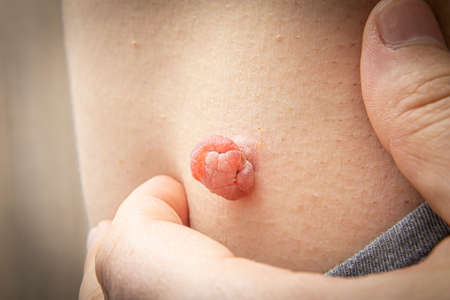
Close Up photo of skin tag or skin mole on a human body swollen and enlarged from the medical dermatologist treatment with liquid nitrogen. Skin mole tag removal. Dermatological beauty treatment
Коллекция по умолчанию
Коллекция по умолчанию
Создать новую
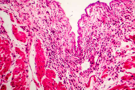
Condyloma acuminatum, also known as genital warts. Light micrograph, photo under microscope
Коллекция по умолчанию
Коллекция по умолчанию
Создать новую
Condyloma acuminatum, also known as genital warts. Light micrograph, photo under microscope
Коллекция по умолчанию
Коллекция по умолчанию
Создать новую
Basal cell cancer Diseased tissue 100x
Коллекция по умолчанию
Коллекция по умолчанию
Создать новую

Ovarian cancer, light micrograph, photo under microscope. Photograph shows a fragment of a cancerous tumor in the female ovary. Selective focus
Коллекция по умолчанию
Коллекция по умолчанию
Создать новую
Bowen's Disease Tumor under the microscope 100x
Коллекция по умолчанию
Коллекция по умолчанию
Создать новую
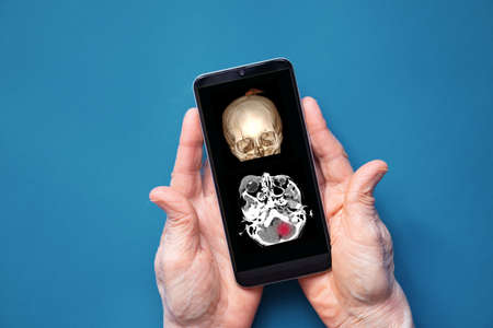
computed tomography of the skull with a crack on the forehead after a fall, skull fracture, computed tomography of 3D skull in the phone in the doctor's hands, mobile application
Коллекция по умолчанию
Коллекция по умолчанию
Создать новую
bacteria cells and virus under the microscope, concept of microbiology, scientific and medical research (3d render)
Коллекция по умолчанию
Коллекция по умолчанию
Создать новую
Doctor demonstrates an X-ray of the male prostate gland, to diagnose the X-ray picture of the prostate, on a white background.
Коллекция по умолчанию
Коллекция по умолчанию
Создать новую

Many moles on the chest of a young man. Check benign moles. The effect of the sun on the skin. The concept of health. Hairy chest, athletic build. Close up.
Коллекция по умолчанию
Коллекция по умолчанию
Создать новую
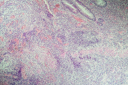
Gastric carcinoma in tissue section 100x
Коллекция по умолчанию
Коллекция по умолчанию
Создать новую
Uterine cancer, light micrograph, photo under microscope
Коллекция по умолчанию
Коллекция по умолчанию
Создать новую

small female breasts close up
Коллекция по умолчанию
Коллекция по умолчанию
Создать новую
Squamous cell carcinoma diseased tissue under the microscope 100x
Коллекция по умолчанию
Коллекция по умолчанию
Создать новую
A macro closeup of a red flesh cell in a body with various sized tumors attached to it - 3D render
Коллекция по умолчанию
Коллекция по умолчанию
Создать новую

Bowen's Disease Tumor under the microscope 100x
Коллекция по умолчанию
Коллекция по умолчанию
Создать новую

Rows of microscope glass slide in the cells
Коллекция по умолчанию
Коллекция по умолчанию
Создать новую

Many different awareness ribbons on red background. World Cancer Day
Коллекция по умолчанию
Коллекция по умолчанию
Создать новую
Salivary gland swollen diseased tissue under the microscope 100x
Коллекция по умолчанию
Коллекция по умолчанию
Создать новую
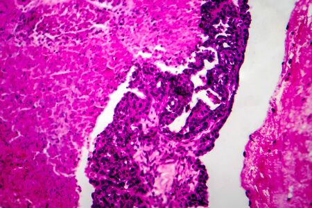
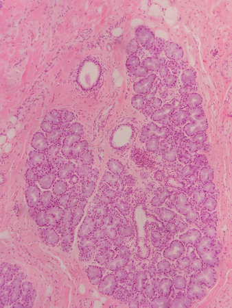
Histopathology of adenocarcinoma of the prostate
Коллекция по умолчанию
Коллекция по умолчанию
Создать новую
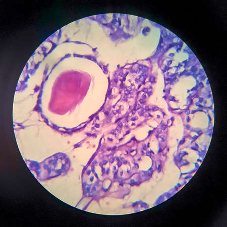
Light micrograph of teratoma, a tumor made up of several different types of tissue, such as hair, teeth, muscle, or bone. Teratoma is typically found in the ovary, testicle, or coccyx
Коллекция по умолчанию
Коллекция по умолчанию
Создать новую
Hairy skin mole. Close up picture of dangerous brown nevus on human skin - melanoma
Коллекция по умолчанию
Коллекция по умолчанию
Создать новую

Red Blood Cells
Коллекция по умолчанию
Коллекция по умолчанию
Создать новую

Composition with different awareness ribbons on purple background. World Cancer Day
Коллекция по умолчанию
Коллекция по умолчанию
Создать новую

Abstract science background- pyloric division of the stomach of the dog. Cell biology
Коллекция по умолчанию
Коллекция по умолчанию
Создать новую
Dermatologist examine birthmark on patient's skin
Коллекция по умолчанию
Коллекция по умолчанию
Создать новую
Bacteria, Bacterial colony, Microbes.
Коллекция по умолчанию
Коллекция по умолчанию
Создать новую
Papillary serous ovarian adenocarcinoma, cancer of ovary, light micrograph, photo under microscope
Коллекция по умолчанию
Коллекция по умолчанию
Создать новую
Histopathology of cirrhosis under the microscope 100x.
Коллекция по умолчанию
Коллекция по умолчанию
Создать новую
Esophageal squamous cell carcinoma, light micrograph, photo under microscope
Коллекция по умолчанию
Коллекция по умолчанию
Создать новую

Different awareness ribbons on color background
Коллекция по умолчанию
Коллекция по умолчанию
Создать новую

Dividing stem cells, 3D illustration. Research and scientific background
Коллекция по умолчанию
Коллекция по умолчанию
Создать новую

Abstract macro image of particles looking like bacteria, macro shot, microbiology theme
Коллекция по умолчанию
Коллекция по умолчанию
Создать новую
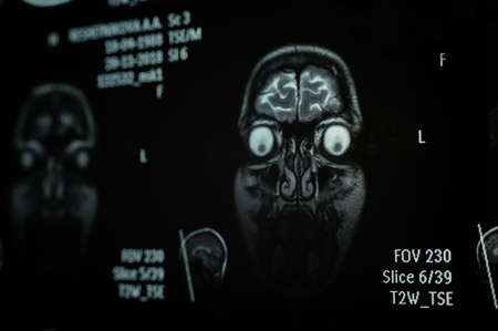
Close-up of magnetic resonance imaging of the human brain
Коллекция по умолчанию
Коллекция по умолчанию
Создать новую
macro of human bloods. microscope view for education physiology .3d microbiology rendering
Коллекция по умолчанию
Коллекция по умолчанию
Создать новую

eczema atopic dermatitis symptom skin texture photo
Коллекция по умолчанию
Коллекция по умолчанию
Создать новую
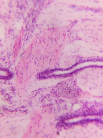
Colon inflammation in Crohn's disease 100x
Коллекция по умолчанию
Коллекция по умолчанию
Создать новую
Bacteria Pediococcus, 3D illustration. Gram-positive cocci, associated with infections in patients with gastrointestinal abnormalities, after abdominal surgery, cause bacteremia, abscess, meningitis
Коллекция по умолчанию
Коллекция по умолчанию
Создать новую

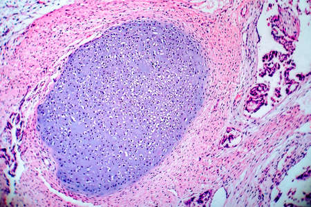
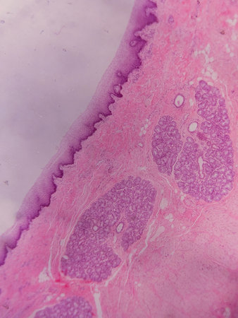
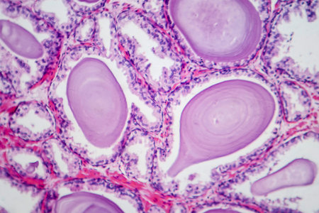
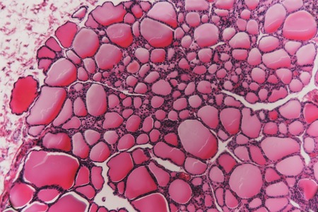
Endemic goiter, light micrograph, abnormal enlargement of the thyroid gland due to dietary iodine deficiency. Photomicrograph shows follicles of varying size, abundant colloid, lymphocytic infiltrate
Коллекция по умолчанию
Коллекция по умолчанию
Создать новую
Painting acrylic paint- abstract drawing. Texture background
Коллекция по умолчанию
Коллекция по умолчанию
Создать новую
Thyroid follicular carcinoma, light micrograph, photo under microscope
Коллекция по умолчанию
Коллекция по умолчанию
Создать новую

Histopathology of interstitial nephritis, light micrograph, photo under microscope
Коллекция по умолчанию
Коллекция по умолчанию
Создать новую
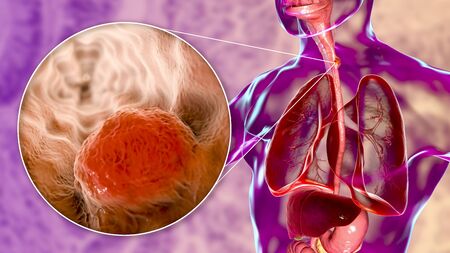
Esophageal cancer, 3D illustration showing malignant tumor in the human esophagus
Коллекция по умолчанию
Коллекция по умолчанию
Создать новую
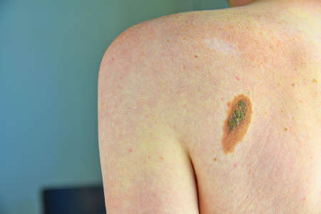
Close-up of a large mole on the body of a caucasian man. macro shot.
Коллекция по умолчанию
Коллекция по умолчанию
Создать новую

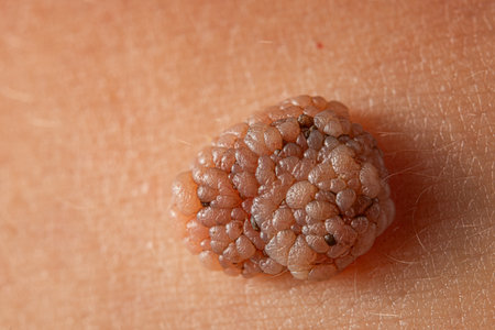
close up of a woman skin focused on a wart
Коллекция по умолчанию
Коллекция по умолчанию
Создать новую
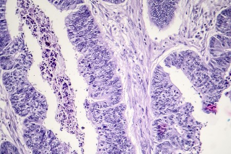
Signet ring cell carcinoma of the stomach, light micrograph, photo under microscope
Коллекция по умолчанию
Коллекция по умолчанию
Создать новую
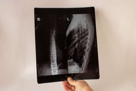
Medical X-ray of the teenagers back.The concept of medical healthcare
Коллекция по умолчанию
Коллекция по умолчанию
Создать новую
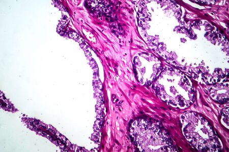
Histopathology of prostate gland hyperplasia, light micrograph, photo under microscope
Коллекция по умолчанию
Коллекция по умолчанию
Создать новую
Pancreas cancer, light micrograph, photo under microscope
Коллекция по умолчанию
Коллекция по умолчанию
Создать новую

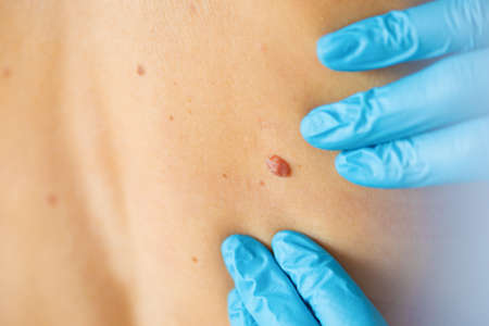
Doctor measuring size of patients pigmented nevus with ruler in clinic closeup. Diagnosis of skin tumors concept
Коллекция по умолчанию
Коллекция по умолчанию
Создать новую
Human liver tissue under microscope view. Histological sample of human liver.
Коллекция по умолчанию
Коллекция по умолчанию
Создать новую
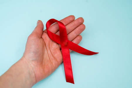
red ribbon on blue background. symbol world aids day.
Коллекция по умолчанию
Коллекция по умолчанию
Создать новую
Gastric carcinoma in tissue section 100x
Коллекция по умолчанию
Коллекция по умолчанию
Создать новую
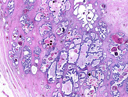
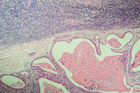
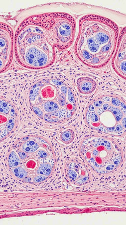
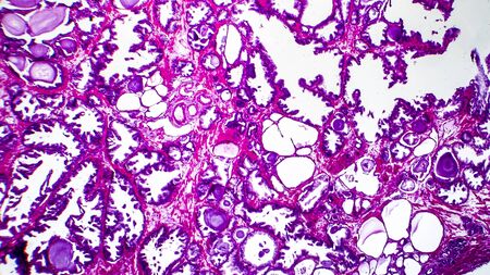
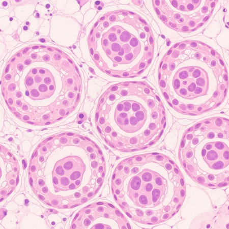
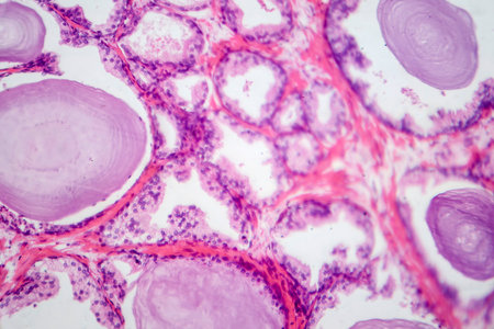
Low magnification of a human prostate gland in a 70-year-old man. The prostate gland appears with dilated alveoli, which contains many corpora amylacea (prostatic concretions) in their lumen. Light microscope micrograph. Hematoxylin & eosin stain.
Коллекция по умолчанию
Коллекция по умолчанию
Создать новую
Histopathology of human liver under microscope view for medical education.
Коллекция по умолчанию
Коллекция по умолчанию
Создать новую

Melanocytic nevus, some of them dyplastic or atypical, on a caucasian man of 37 years old from Spain
Коллекция по умолчанию
Коллекция по умолчанию
Создать новую
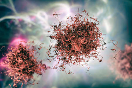
Cancer cell growth uncontrollably over tissue, Tumor infection cells and spreading, Invasive inflammation metastasis cancerous. reproduce by duplicating, cells expanding, Melanoma
Коллекция по умолчанию
Коллекция по умолчанию
Создать новую
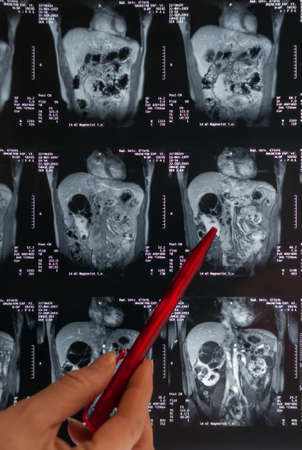
Magnetic resonance imaging, MRI, computed tomography, x-ray image. Area of ​​the pelvis with kidneys infected with tumors
Коллекция по умолчанию
Коллекция по умолчанию
Создать новую
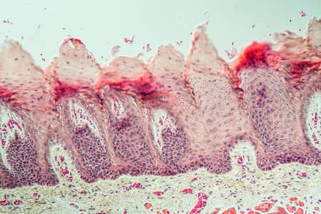
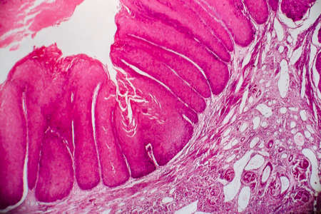
Tongue Tissue with taste buds across 200x
Коллекция по умолчанию
Коллекция по умолчанию
Создать новую
Breast cancer, light micrograph, photo under microscope
Коллекция по умолчанию
Коллекция по умолчанию
Создать новую
Photomicrograph showing histological features of benign prostatic hyperplasia. Enlarged prostate gland with nodular proliferation of glandular and stromal components.
Коллекция по умолчанию
Коллекция по умолчанию
Создать новую
Breast cancer, light micrograph, photo under microscope
Коллекция по умолчанию
Коллекция по умолчанию
Создать новую
A longitudinal section of human spinal ganglion cells under the microscope.
Коллекция по умолчанию
Коллекция по умолчанию
Создать новую
Raw beef lungs on white backgroundу Top view
Коллекция по умолчанию
Коллекция по умолчанию
Создать новую
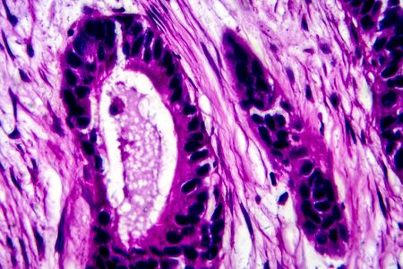
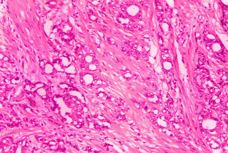
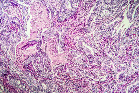
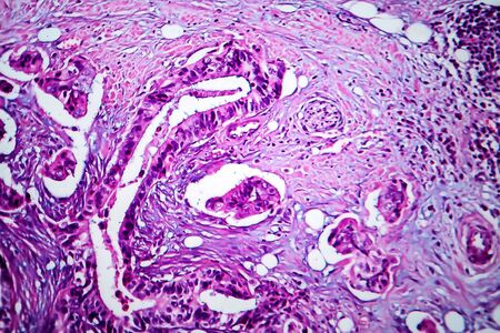
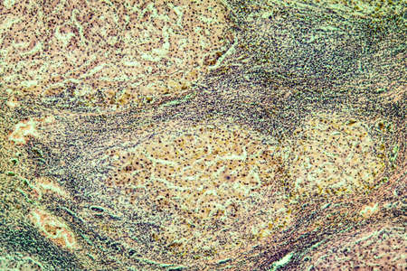
A microscopic view of tissue with pink and purple staining, showing cellular structures and patterns
Коллекция по умолчанию
Коллекция по умолчанию
Создать новую
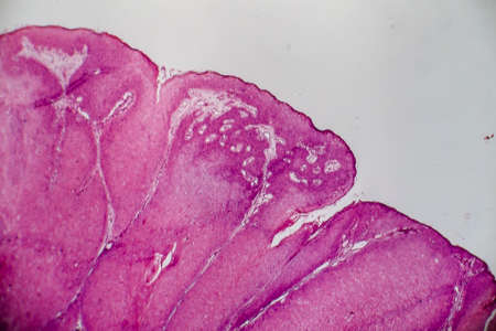
Showing Light micrograph of the Adrenal gland and Urinary bladder human under the microscope for education in the laboratory.
Коллекция по умолчанию
Коллекция по умолчанию
Создать новую

Breast cancer symbol. Three pink ribbons
Коллекция по умолчанию
Коллекция по умолчанию
Создать новую
Cancer cells abstract background. 3d illustration
Коллекция по умолчанию
Коллекция по умолчанию
Создать новую
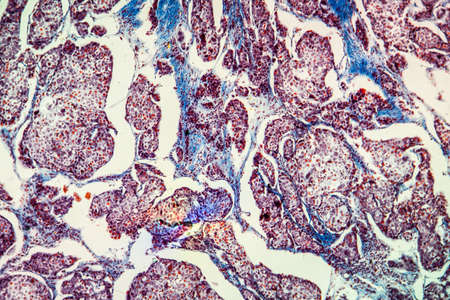
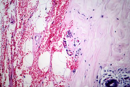
Liver cirrhosis tissue affected 100x after alcohol abuse
Коллекция по умолчанию
Коллекция по умолчанию
Создать новую
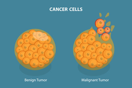
3D Isometric Flat Vector Illustration of Cancer Cells, Tumor Development
Коллекция по умолчанию
Коллекция по умолчанию
Создать новую
Photomicrograph showing histological features of benign prostatic hyperplasia. Enlarged prostate gland with nodular proliferation of glandular and stromal components.
Коллекция по умолчанию
Коллекция по умолчанию
Создать новую
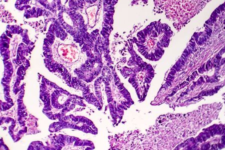
Prostate cancer, light micrograph, photo under microscope
Коллекция по умолчанию
Коллекция по умолчанию
Создать новую
Differentiated intestinal adenocarcinoma, light micrograph, photo under microscope
Коллекция по умолчанию
Коллекция по умолчанию
Создать новую

Seamless surface dividing cells on white background closeup. A high resolution.
Коллекция по умолчанию
Коллекция по умолчанию
Создать новую

Bladder cat- cell nature background. Abstract- photo macro sections with high magnification with light microscope
Коллекция по умолчанию
Коллекция по умолчанию
Создать новую
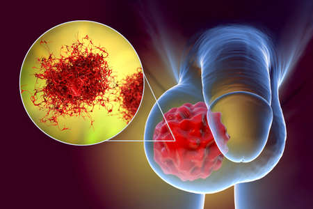
Testicular cancer, testicular seminoma, 3D illustration and light micrograph. Malignant tumor of the testis
Коллекция по умолчанию
Коллекция по умолчанию
Создать новую
Yellow ribbon on purple background
Коллекция по умолчанию
Коллекция по умолчанию
Создать новую

Orange awareness ribbon on yellow background. Kidney cancer concept
Коллекция по умолчанию
Коллекция по умолчанию
Создать новую

Thyroid follicular carcinoma, light micrograph, photo under microscope
Коллекция по умолчанию
Коллекция по умолчанию
Создать новую
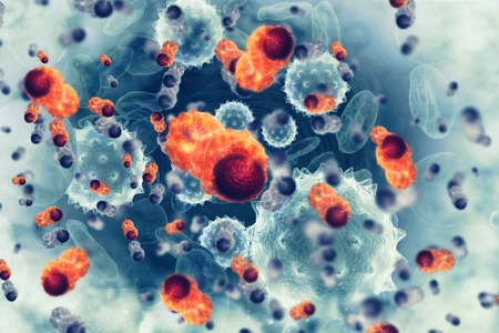
Cancer cells.3d illustration
Коллекция по умолчанию
Коллекция по умолчанию
Создать новую
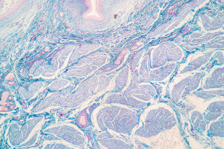
Abstract science- biology tissue thyroid of dog. Medical and biological tissue prepared microscope slide. Educational material for the study and treatment of animals.
Коллекция по умолчанию
Коллекция по умолчанию
Создать новую
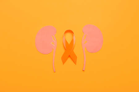
Awareness ribbon and paper kidneys on orange background
Коллекция по умолчанию
Коллекция по умолчанию
Создать новую

Chronic pyelonephritis, light micrograph, photo under microscope. High magnification
Коллекция по умолчанию
Коллекция по умолчанию
Создать новую
Cancer cell, malignant tumor cell, 3D illustration
Коллекция по умолчанию
Коллекция по умолчанию
Создать новую
Histopathology of interstitial nephritis, light micrograph, photo under microscope. High magnification
Коллекция по умолчанию
Коллекция по умолчанию
Создать новую
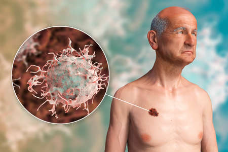
Melanoma, a cancer developing from pigment-containing cells melanocytes, 3D illustration showing melanoma on the man's trunk and close-up view of cancer invasion
Коллекция по умолчанию
Коллекция по умолчанию
Создать новую
Migrating cancer cell
Коллекция по умолчанию
Коллекция по умолчанию
Создать новую
Bladder cancer, light micrograph, photo under microscope. High magnification
Коллекция по умолчанию
Коллекция по умолчанию
Создать новую
Anatomy and Histological Ovary, Testis and Sperm human cells under microscope.
Коллекция по умолчанию
Коллекция по умолчанию
Создать новую
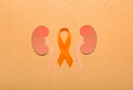
Orange awareness ribbon and paper kidneys on beige background
Коллекция по умолчанию
Коллекция по умолчанию
Создать новую
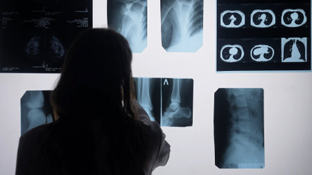
Doctor analyzing X Ray images for medical diagnosis
Коллекция по умолчанию
Коллекция по умолчанию
Создать новую
Legion-Media
Создайте свои проекты на основе качественных стоковых фотографий и видео.


Copyright © Legion-Media.